

In a recent post, I discussed how it’s now possible to coax neurons in the central nervous system to express new receptors, allowing them to be remotely controlled by a previously inert drug. Chemogenetics could be applicable for a wide range of ailments in neurology, from epilepsy to neuropathic pain: anything that can be alleviated by increasing or decreasing the activity of a subset of neurons.
However, chemogenetics can only help when the required effector neurons exist. In late-stage Parkinson’s disease (PD), for example, dopaminergic neurons are so depleted that conventional treatments that increase the activity of spared neurons fail. Chemogenetics couldn’t help at this stage. What if we could instead replace neurons?
There’s a lot of excitement right now about neuron replacement therapies for the CNS. In this primer, I’ll give a bird’s-eye view of where the field is, what the bottlenecks are, and whether neuron replacement therapies could be accelerated with AI. I’ll focus on the particularly illustrative case of neuron replacement therapies for Parkinson’s disease (PD), which have been around for almost 4 decades, and which show in some detail how the field has stumbled and evolved. I’ll show that there are a number of bottlenecks in neuron replacement therapies that could be ameliorated through AI, but that we’ll need new datasets, infrastructure, and regulation to reach that point.
But first, let’s explore what in my view is one of the more promising applications: epilepsy.
Where we are now
A prime example of a CNS neuron replacement therapy is Neurona, a company building a treatment for epilepsy based on embryonic stem cells (hESC) called rezanecel. Epilepsy occurs when there’s runaway excitation in a given brain area. As I covered in the essay on chemogenetics, treatments that decrease excitation globally have deleterious side effects, and a sizable fraction of epilepsy patients cannot get it under control. A targeted therapy should decrease local excitation or increase local inhibition near the epileptic focus; Neurona increases local inhibition.
Time-lapse of interneurons shmoving through the mouse MGE. Upsampled from Lepiemme et al. (2021).
Neurona’s treatment is made from a human embryonic stem cell bank. The cells are coaxed into taking on the identity of GABAergic interneurons as they appear during development. One of the remarkable things about the development of the nervous system is that inhibitory cells are born in a different physical location than excitatory cells: cells in the medial ganglionic eminence (MGE), which eventually become PV+ (parvalbumin) and SOM+ (somatostatin) cells, migrate over long distances—hundreds of times their body lengths—to reach their final destination. Coaxing stem cells into this intermediate neuronal state—post-mitotic, but still migratory—means the cells can move from their original location and integrate into the local network: it is in their very nature. When they’re injected into the long axis of the hippocampus with epilepsy, they put a brake on local activity.
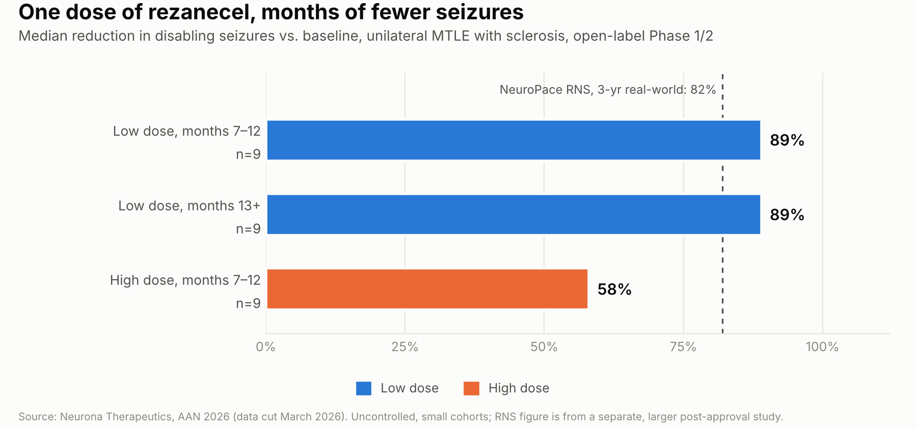
The early results look promising: in an open-label phase 1/2 clinical trial, low-dose rezanecel decreased the incidence of disabling seizures by 89% (N=9) in unilateral temporal lobe epilepsy. Is that a lot? Yes! By contrast, one brain interface, the NeuroPace responsive neurostimulator, reported an 82% median seizure reduction in its 3-year post-approval study. A cell therapy like rezanecel could make a sizable dent in the disease burden of epilepsy, pending the results of the Phase 3 clinical trial which will start recruiting in late 2026.
Neurona is not the only late-stage CNS neuron replacement therapy that is showing promise for a neurological disorder. BlueRock has a neuron replacement therapy for Parkinson’s disease called bemdaneprocel; its estimated primary completion for its phase 3 is 2027. They have the backing of the pharma giant Bayer, which acquired them in 2019. Another notable example is Amchepry (raguneprocel) from Sumitomo Pharma, also for PD: they received conditional approval in Japan in March 2026, which means that they can manufacture and sell it for the next 7 years within the Japanese healthcare system, on the basis of a Phase I/II study.
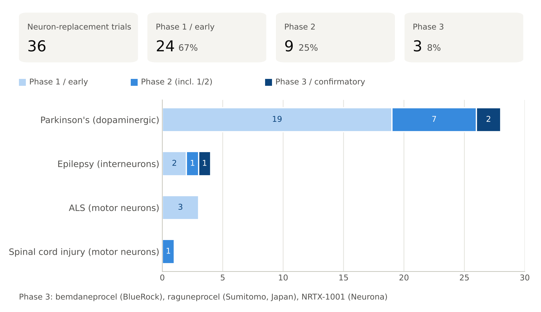
I had Claude fetch data from clinicaltrials.gov to characterize clinical trials testing neuron replacement. PD and epilepsy form the bulk of these efforts, presumably because of their relatively well-characterized, cell-type-specific disease mechanisms that can be straightforwardly addressed through neuronal therapies. One could, however, imagine a much larger range of potential neurological disorders addressed by neuron replacement therapies. Given the sci-fi nature of the therapies, and their substantial promise, one might reasonably ask what’s taking so long?
How we got here
It may seem like a naive question to ask why it took so long to get here. If neuron replacement therapies are approved in the US in the next couple of years, that would mean approximately 40 years between first-in-human tests attempting neural replacement and approval. Was some of this wait avoidable? It’s a little hard to appreciate just how different the therapies of today are from the pioneer therapies of yesteryear. Let’s go back to the start of it all, in PD, where the bulk of clinical trials are, and where the historical record has been well documented.
First-generation products: fetal tissue grafts
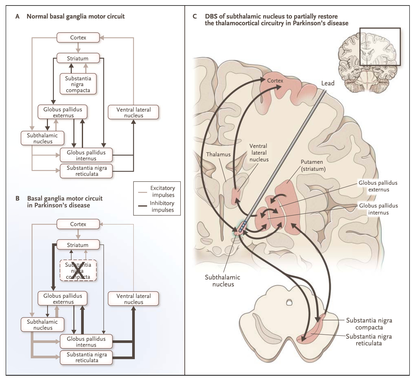
Parkinson’s disease (PD) is a neurodegenerative disorder that causes the progressive loss of dopamine-releasing neurons in the midbrain. Many treatments for PD coax the remaining cells or other nodes in the basal ganglia to compensate for this loss, whether that’s using L-DOPA to keep a constant supply of the dopamine precursor, or deep brain stimulation to electrically stimulate subthalamic nuclei that are part of the same circuit. Since the lost dopaminergic cells project to the putamen, it would stand to reason that directly injecting cells that produce dopamine into the putamen would alleviate some of these symptoms.
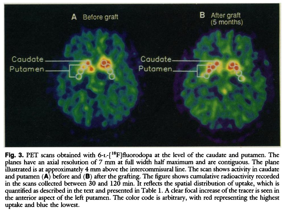
In 1987, Lindvall & Bjorklund at the University of Lund in Sweden first grafted fetal dopaminergic neuroblasts (immature precursor cells to neurons) in PD patients. In 1990, injecting into the putamen, they showed that the grafts survive; and in 1999, they showed that a graft into the putamen restored dopamine release, allowing the patient to come off L-dopa for years.
These early positive results were later countered by more pessimistic outcomes in large-scale clinical trials. In 2001, a larger clinical trial published in the New England Journal of Medicine found no overall benefit on the primary outcome compared to the sham control (Freed et al. 2001). Worryingly, roughly 15% of patients suffered dyskinesias off medication, involuntary movements that negatively affect quality of life. Another sham-controlled study in 2003 found that more than half of patients who received cell grafts suffered dyskinesias (Olanow et al. 2003).
It’s important to understand that cell therapies are a different beast entirely than, say, small molecules: sourcing and manufacturing are highly non-trivial. In the case of fetal transplants, it requires the dissection of fetal tissue at a particular stage of development and the isolation of the right cell type; oftentimes, cells from multiple fetuses must be pooled together to gather the millions of cells necessary for transplantation. The grafted cells thus carry different genetic identities from the host and from each other, and may include different cell fates.
One theory of the early dyskinesias is that they were caused by what one might call a manufacturing defect: the batches of putative dopaminergic cells may have contained undesirable serotonergic neurons. These serotonergic neurons can take up dopamine, convert and package it, and release it as a false transmitter. They don’t have the native autoreceptors of dopamine neurons, meaning they can cause unregulated, pulsatile dopamine release. This is by no means the only theory, however: others have instead blamed local immune reaction, which is hard to prevent with cells from multiple donors.
Many years later, the TransEuro collaboration ran a clinical trial in which they more tightly controlled the source of the cells, the dissection, and put patients on 12 months of triple immunosuppression. The collaboration suffered many delays in procuring the right cells: only 11 patients were grafted. It is highly non-trivial to isolate millions of cells under rigorous QC while eliminating contaminants when the raw supply is extremely limited.
While no major dyskinesias were found, only one patient ultimately reached near-normal dopamine uptake as measured through PET. The authors of the collaboration (Barker et al. 2025) blamed in part supply issues for this negative result: using tighter controls forced them to pool tissues from different gestational ages and to use less of them.
> variation in the gestational age of the donors for each patient, the shortage of tissue available for each patient and the limited possibilities for standardization and quality control inherently lead to variation in outcome — Barker et al. (2025)
While early experiments and follow-ups revealed that there was a glimmer of hope in neural transplants, it also made it clear that the fetal source was a bottleneck. In addition to their technical drawbacks, quality control issues, and limited supply, fetal transplants are highly contentious politically: such a study could not have been funded by the NIH under current rules. The TransEuro collaboration concluded that the future was instead in stem-cell sources.
Second-generation products: allogeneic hESCs and iPSCs
A far more scalable source of dopamine neurons are stem cells. Human embryonic stem cells (hESCs) are derived from the extra embryos generated during IVF1. Induced pluripotent stem cells (iPSCs) are instead derived from skin or blood cells: they shed their aged phenotype and become pluripotent after exposure to the 4 transcription factors discovered by Yamanaka in 2006, for which he was awarded the Nobel Prize in 20122.
Stem cells obtained from banks can then be coaxed, using small molecules and growth factors that mimic developmental signals, to become dopaminergic cell progenitors. Figuring out how to get just the right cell identity is highly non-trivial. Building on the most successful fetal graft studies, Kriks et al. (2011) focused on coaxing hPSCs into a dopaminergic neuron identity by mimicking the development environment of the midbrain floor plate, where dopaminergic neurons are born. They also needed to fine-tune the protocol so that cells are not so late in the development process that they don’t engraft, but not so early that they overgrow. Their protocol became the basis for BlueRock’s product; Amchepry, the PD cell therapy from Sumitomo, uses a closely related protocol.
hESCs and iPSCs are far more scalable cell products than fetal grafts: one master cell bank can make lots for many patients. Furthermore, one can tightly sort for dopamine progenitors or tightly define differentiation. But it’s important to understand just how tightly these cell products must be manufactured: in animal studies, as few as 100 undifferentiated stem cells (0.01%) left in a dose can form nightmare-inducing teratomas, tumors that chaotically grow skin, hair, and teeth.
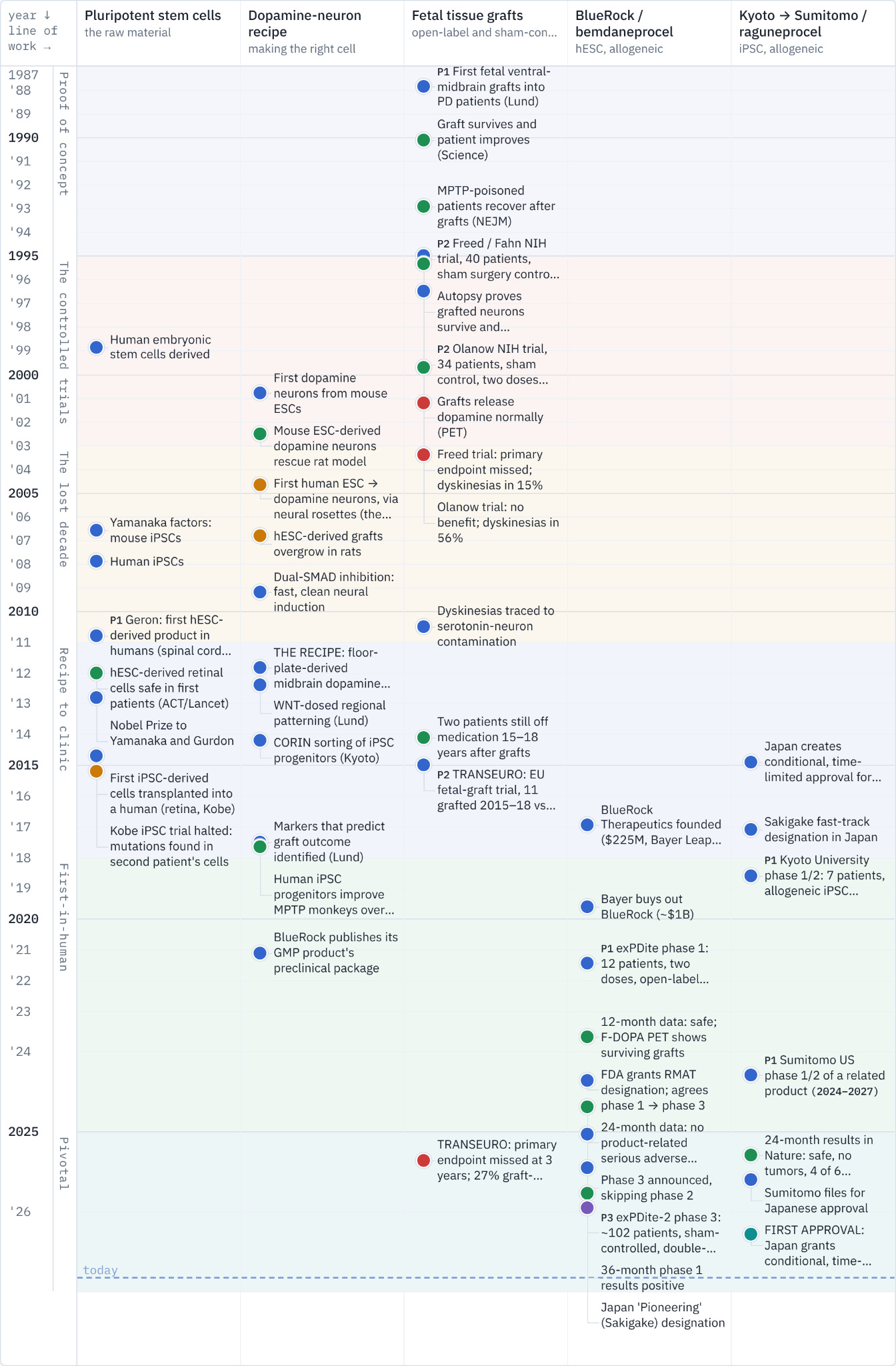
So between 1987 and 2011, fundamental scientific discoveries allowed us to source neuron precursors and differentiate them far more reliably, and we also learned from early experiments important lessons about the importance of immunosuppression. This helped pave the way for the results reported recently.
Does it work?
The results of PD dopaminergic cell precursor injections were reported back-to-back in Nature in April 2025: BlueRock’s product, derived from hESCs, and Sumitomo’s product, derived from iPSCs. Between 900k to 5.5M cells, depending on the experimental arm, were bilaterally injected into the putamen of patients; no patients suffered serious adverse events. Grafts appeared to survive well and take up F-DOPA in PET scans.
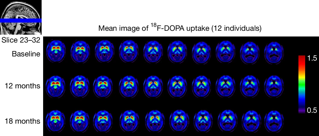
The Kyoto trial, the basis for Amchepry, reported that 4 out of 6 patients improved when they were off medication, with a mean change of -9.5 points on a motor scale (20%) where lower is better. The BlueRock trial showed a headline result of -23 points (48%) at 18 months and -17.9 points (37%) at 36 months at the highest dose. In both cases, trials were very small and open-label, so one shouldn’t read too much into the exact numbers.
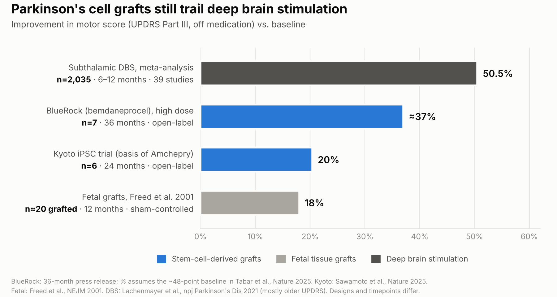
Is that a lot? To give a point of reference, in a recent meta-analysis (Lachenmayer et al. 2021), patients in deep brain stimulation trials were found to improve on a similar scale by 50%. DBS, like neuron replacement therapies, requires brain surgery; it is proven, and it is cheaper than the neuron replacement therapies by a factor of at least 2X3. So why should we be excited about PD neuron replacement therapy?
Cell therapies are very flexible, and there is no telling what the upper bound is for efficacy. Consider CAR-T (chimeric antigen receptor T cells), for example, which reprogram the immune system to recognize and kill cancer cells. First-generation CAR-Ts, starting in 1989, did not persist in patients and thus had little effect. It was only when they were edited with a costimulatory domain that the cells could expand and survive. In 2014, a trial in children and adults with relapsed acute lymphoblastic leukemia reported complete remission in 27 of 30 patients (90%). The FDA approved Kymriah in 2017, and it is now being expanded beyond cancer for autoimmune disease. Similarly, if one built upon the existing encouraging results in PD neuron replacement therapy, we might have a highly efficacious therapy available.
The garden of forking paths
We can think of many, many ways in which these technologies might be improved. For example, dopaminergic cell differentiation in grafts is quite poor: A systematic review (Comini & Dowd 2025) found a median differentiation of 3% into dopaminergic neurons across a range of pre-clinical trials. One could engineer cells to better become integrated into the environment by responding to local cues, or co-injecting with glial cells to coax cells into the right fate.
As long as one is gene-editing the cells, one can make them remotely controllable with DREADDs, as I covered in my previous essay, so they gain the fine-tuning capability of DBS. One could edit them for immune evasion so they become hypoimmune—though that comes with its own disadvantages, including cancer risk. One could instead derive the iPSCs from the same patient—autologously—so patients don’t need to be on immunosuppressants, as Aspen Neuroscience is proposing. If one wants to promote long-term survival, one might also want to knock out the α-synuclein gene to prevent protein aggregation, Lewy bodies and neurodegeneration.
And what about delivery? The TransEuro collaboration used different versions of the implantation device that seemed to affect outcomes: these are physical devices which, when poorly designed, can squish cells, unevenly dispense liquid and solids (think of a jar of natural peanut butter that’s all oil at the top and all solids at the bottom), and cause backflow. And would it not be better to figure out a way to systemically inject cells and have them distribute themselves to the right location in the brain?
The PD example illustrates that as a platform, CNS cell therapies have a much higher ceiling than what they have been able to reach. A half dozen or so tweaks are named here; a more thorough review would undoubtedly reveal many more. If we could speedrun their development, neuron replacement therapies could act as a new pillar modality. But these ideas demand biological verification. Could we accelerate these developments using AI, or will we have to wait decades to get to truly futuristic therapies?
So what’s taking so long?
It took two decades of open-ended science to get to a point where we had the knowledge to start running preclinical research and clinical trials. Understanding how this process unfolded could help us prepare for future therapies. Broadly, we can think of these epochs:
Early promise and failure of fetal grafts (1987-2003): Fetal tissue transplants show promise, but NIH-funded sham-controlled trials miss their primary endpoint
Lost decade (2003-2011): while no major PD cell therapy trials are running, scientific breakthroughs lead to iPSCs and the floor-plate protocol for midbrain dopamine neurons
The pre-clinical period (2011-2018): Groups build GMP lines to transform the small-scale dopamine neuron protocol into a manufacturable product; new dopamine neurons are tested for tumorigenicity (6-9 months) and for efficacy in MPTP monkeys (24 months).
First-in-human (2018-2025): Several first-in-human studies are run: Kyoto (2018+), BlueRock (2021+), Aspen (2024+). Recruitment takes 1-2 years, and readouts are 18-24 months later.
2025+: Pivotal and onwards
Some delays are irreducible: one could argue that open-ended scientific inquiry—the period from 1987 to 2011—is difficult to systematically accelerate. Clinical trials add fixed delays: grafts take time to mature, and readouts occur when grafts have matured, typically at 18-24 months.
Some delays are partly driven by policy: many PD cell therapy trials are sham-controlled. That means patients in the placebo arm get small holes drilled into their skull, but don’t receive cell injection. Early trials revealed a sizable placebo effect, and it has become common practice to have sham-surgery-controlled clinical trials. Sham surgery likely delays recruitment, because few want to be enrolled in a trial where one has a sizable chance of receiving a sham therapy; one patient advocacy group survey showed only 17% of respondents would volunteer for a trial involving a sham surgery arm (Cohen et al. 2010).
There are many potential solutions: leveraging platform trials that share a control arm, using synthetic controls, replacing the sham surgery arm with an active control like DBS, using a cross-over design where all patients eventually receive the therapy, and approving medicines conditionally based on open-label studies followed by post-market studies.
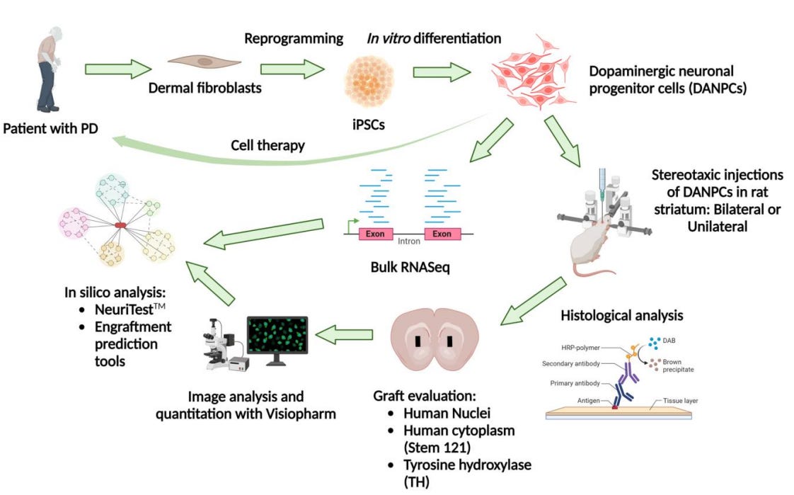
Some delays could conceivably have been accelerated by AI. Manufacturing cell lines at scale is one example of that, and it’s perhaps best illustrated by one of the upstart entrants in the space, Aspen Neuroscience, which is building autologously derived implants that don’t require immune suppression. This fascinating deck goes through the process by which they derive iPSCs and differentiate them into dopaminergic cell precursors. There are dozens of steps involved in that process, many of them involving computer vision for QC, reagents, changing media, putting transcription factors in, etc. These are complex recipes derived from the acquired wisdom of hundreds of grad students.
The manufacturing process for iPSC-derived products is generally estimated, industry-wide, to take 6 months and cost $0.5-1M (Doss & Sachinidis 2019). Could I see this be accelerated and made radically cheaper with lab robotics and an AI scientist reading the entire literature and closely monitoring every step of the process, the same way that auto-GPT attempts to automate ML research? Absolutely. In fact, there’s a project from Japan’s CiRA foundation that aims to do just that for deriving iPSC cell lines.
As we ponder future neuron replacement therapies, we will run into technical problems that are not addressable by AI models today, but that we could mold into AI-shaped problems. Consider graft survival and differentiation: it is not immediately obvious how one might predict which edits to neuron precursors will make them more or less likely to implant correctly in an aged brain. Neuronal survival and differentiation is canonically a long-horizon problem: it can take years for cells to integrate themselves into the local environment. Where would you even get the data for that? Could one transform this into an AI-shaped problem by screening on aged mice? Ex-vivo brains? Decedent models?
It has taken 39 years for neuron replacement therapies to go from experiment to conditional approval. We know that the first treatments that will come online will not be perfect; but the ceiling for a cell therapy could potentially be very high. In a peri-AGI world where making (virtual) cell edits is a matter of a couple of clicks, verification and running clinical trials at scale are increasingly pressing. AI will not magically solve disease, but addressing AI-shaped problems, molding non-AI-shaped problems to make them more AI-shaped, and solving policy bottlenecks might get us there.
Thanks to Alex Plesa for feedback on this draft.
While these have also been the source of controversy and the policies have swung with administrations, embryonic stem cells are far less supply-constrained: a single embryo can be used to derive an essentially eternal cell line.
iPSCs’ main theoretical advantage over hESCs is that they can be made autologous, where a patient’s own cells serve as a basis for the therapy; however, they are less of a blank slate than hESCs, leaving open questions about aberrant methylation, mutations, and residual memory. We’ll lump the two technologies together for the purpose of this already long blog post.
Amchepry retails for ¥55.3M, or about $350k, compared to an all-in 5-year cost for DBS in the US of $180k
Thank you Patrick, very thorough analysis!
The TransEuro example makes me want delivery details kept alongside cell-manufacturing data. Device and surgical site changed together, and the Lund patients had less advanced disease. Otherwise a model might credit the cell preparation for a difference it cannot separate from delivery or patient selection.